|
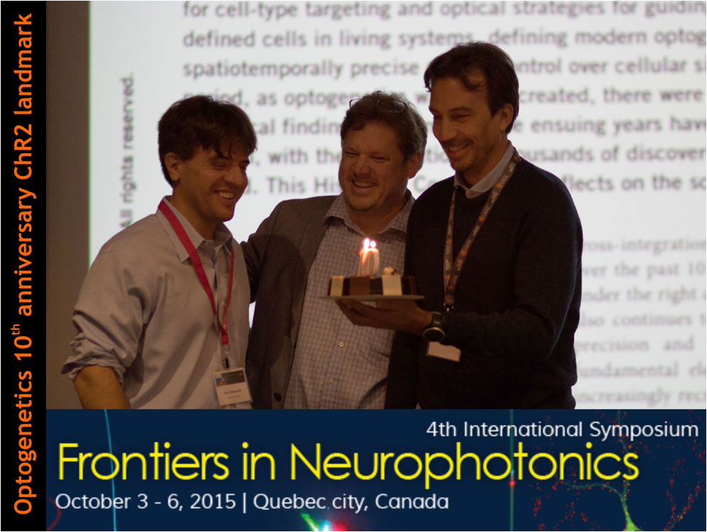
Synapses, brain cells, and neural networks are key elements to understanding the age-old question, “What makes us tick?” While there is always a need for increased detail while imaging the tiny events at synapses, there is also a requirement to understand the population of neurons that interact in complicated networks. This range of scales that must be explored simultaneously is what makes the brain such a difficult organ to decode. With the application of novel super-resolution imaging techniques, the development of innovative optical devices as well as advancements in the chemical engineering of photosensitive proteins, optical methods provide unparalleled functionality for investigating the brain. October 3rd marked the beginning of the fourth biennial Frontiers in Neurophotonics conference, held in Quebec City’s Musée de la Civilisation, offering a gorgeous view of the city’s seaport and the docked Queen Mary 2. Created jointly in 2008 by Université Laval and Université de Bordeaux, this biennial interdisciplinary symposium has successfully, through its different editions, allowed experts to share on leading-edge photonics technologies used to understand brain function and structure. Although it was a small and tight-knit conference, it welcomed some of the world’s leading researchers in neurophotonics, providing an elite series of presentations and discussions. With more than a hundred participants at every edition, it has become the leading neurophotonics forum. The broad range of topics presented at this year’s Frontiers in Neurophotonics conference included novel techniques in acquiring, processing, and analyzing image datasets, new microscopic and fiber-optic tools to probe and manipulate neurons, and impressive advancements in the design of integral photosensitive proteins. Yet one of the hot topics of this symposium, which still remains a major issue for experts, was indisputably the quest to solve the trade-off between space and time resolution in living biological systems. Indeed, super-resolution imaging has become more and more popular in the last decade but is still often limited to fixed tissues. Thus, super-resolution microscopy has been mostly restricted to very simple biological systems and hardly combined with other techniques. Yet, recent advances have made easier the use of super-resolution techniques in more physiological and intact systems. The combination of super-resolution with other known techniques now allows the investigation of the nanoscopic molecular organization of single spine in its characterized environment. By doing STORM imaging following some electrophysiological recordings and confocal imaging, István Katona and his team (IEM, Budapest) elegantly showed how it is now possible to assess endocannabinoid receptors’ precise localization in axon terminals of electrophysiologically and anatomically characterized neurons. Tying the functional and structural connectivity of circuits has been a growing endeavor. Rainer Friedrich (FMI, Basel) presented serial block face-scanning electron microscopy and tedious three-dimensional reconstruction of the olfactory circuit of zebrafish following live calcium imaging in the same tissue exposed to odors, allowing detailed investigation of neuronal computation based on dynamic and structural measurements. Monitoring how synaptic plasticity-inducing stimuli impact the long-term fate of individual synapses has been quite challenging. Thomas Oertner (ZMNH, Hamburg) combined the control and sensing components of optogenetics in organotypic hippocampal slices to describe the fate, over seven days, of individual synapses following depression or potentiation protocols. These different advances in combining optical methods with high resolution will be revealed to be particularly important in the next years to decipher synaptic signaling and remodeling. Besides impressive software unveiling, there were also some optical-path manipulation techniques revealed to increase both the ease of image acquisition and the quality of deep tissue imaging. One example of this came from Jean Baptiste Sibarita and his team (Université de Bordeaux, France), who demonstrated an impressive advancement in the technique of selective plane illumination microscopy (SPIM) by reducing the number of objectives required to perform the procedure from two to one, thus coining the term single objective SPIM (soSPIM). They accomplished this by the use of micromachined mirror-coupled microwells which allowed the single objective to illuminate and collect via different light pathways. Using this technique they showed the capability to image from whole drosophila embryos down to the single cell level, with single molecule detection. Furthermore, as a great example of how all science continues to combine and mix among subgenres, Jérôme Mertz (Boston University, USA) presented how optical tools from one category of research can be used to improve another. He presented experimental demonstrations of an improved, neuroscience-adapted, version of the long-serving method of adaptive optics from the field of astronomy, this time, without the need for guide stars. Using this technique, it should be possible to correct for some of the earlier unavoidable aberrations that arise from imaging through scattering tissue. As an alternative to imaging, probing neurons optically with fiber optics can offer minimally invasive means to reach specific neurons in deep brain structures. Yves De Koninck (Université Laval, Canada) showed novel fiber-optic based probes capable of providing electrical field recording combined with the ability to optically stimulate and sense single neurons in behaving animals. Building on that, he showed some exciting future possibilities using electro-conducting glasses to allow electrolyte-free micro-optrodes for long-term recording. Getting rid of wires will eventually push the limits of our capacity to relate brain activity with complex behavior. Benoit Gosselin (Université Laval, Canada) is developing miniature wireless transmission devices, which are implanted on a live mouse head, that contain an array of electrical probes and LEDs to both stimulate and record activity in behaving animals. As smaller is better in this case, they are further reducing the size of the implants to reduce the load. Auspiciously, the conference also delved deeper into the chemistry behind the scenes. Integral to imaging, understanding, and manipulating neurons of any type is the development of light-sensitive proteins. On this topic, Takeharu Nagai (ISIR, Osaka) presented his now vast palette of nanolantern probes, which take advantage of the bioluminescence resonance energy transfer (BRET) from enhanced luciferase to a fluorescent protein. Since it doesn’t require light, these new probes allow rapid and nontoxic imaging for many hours in cells, in addition to being suitable for dual imaging/optogenetic experiments. Focusing on label-free optical methods, Benjamin Rappaz (EPFL, Switzerland) worked on the development and optimization of digital holographic imaging. By measuring the water fluxes associated with ion fluxes through the membrane, this technique allows the noninvasive fast screening of GABA channel activity. Robert Campbell (University of Alberta, Canada) also gave an update on his teams’ recent strides in creating photosensitive proteins that absorb light further into the red end of the spectrum — as “redder is better” to probe and excite deeper into the tissue, as well as their advancements in reporters for membrane potential, neurotransmitters, and calcium ions. While observing and controlling neurons with optics is providing new knowledge on neuronal functions, many are already looking forward to use light to not only observe but also control various intracellular signaling processes. Some stunning experiments from Casper Hoogenraad (Utrecht, Holland) showed how it is now possible to control the intracellular organelles transport and positioning by simply shining light on specific areas of the cell. In the final hours of Frontiers in Neurophotonics, there was no better way to complete an already outstanding meeting than to have a keynote presentation by Karl Deisseroth (Stanford University, USA), but only after the organizers offered him a special “birthday cake” to celebrate the tenth anniversary of his demonstration with Ed Boyden of the use of light to drive action potentials within neurons expressing a foreign gene borrowed from a unicellular algae. Karl presented impressive results his team has recently obtained in their quest to better understand fear memory pathways in live behaving mice, as well as their progression in developing fast and bistable neuronal spike inhibiting opsins. From improved microscopy techniques to novel optical fibers, all the way to enhanced photosensitive protein engineering, Frontiers in Neurophotonics unveiled exciting discoveries and developments that will continue to influence the field of neuroscience for years to come. Perhaps even more exciting is the realization that the field of neurophotonics is still in its infancy and that it holds a very bright future. The next installment (5th edition) of this conference will be held in 2017 in Quebec’s sister city, Bordeaux, France, and is expected with much anticipation. |


Neurophotonics
Brain
Neurons
Proteins
Super resolution
Tissue optics
Tissues


